What Is Revision Rhinoplasty?
Revision rhinoplasty is generally more complex than primary rhinoplasty because previous surgery
may have weakened or damaged the nasal framework and the soft tissues covering it.
Successful revision surgery requires a careful assessment of why the previous procedure was unsuccessful and of the nose’s current condition.
Cartilage and soft-tissue damage may be more extensive than anticipated, and previously placed materials may be discovered during surgery.
For these reasons, revision rhinoplasty requires a surgeon with substantial experience in complex nasal anatomy and reconstruction.
At Kobijou Rhinoplasty Clinic, we determine the cause of the problem and plan treatment through a physical examination, nasal endoscopy,
and computed tomography (CT). We draw on our extensive experience in revision rhinoplasty to achieve the best possible outcome.
Revision rhinoplasty is generally more complex than primary rhinoplasty because previous surgery
may have weakened or damaged the nasal framework and the soft tissues covering it.
Successful revision surgery requires a careful assessment
of why the previous procedure was unsuccessful and of
the nose’s current condition. Cartilage and soft-tissue damage
may be more extensive than anticipated, and previously
placed materials may be discovered during surgery.
For these reasons, revision rhinoplasty requires a surgeon
with substantial experience in complex nasal anatomy
and reconstruction. At Kobijou Rhinoplasty Clinic,
we determine the cause of the problem and plan
treatment through a physical examination, nasal endoscopy,
and computed tomography (CT).
We draw on our extensive experience in revision rhinoplasty to achieve the best possible outcome.
Timing of Revision Rhinoplasty
Revision surgery is generally considered at least 6 months after the previous procedure, once the tissues have stabilized.
The appropriate timing may vary depending on the patient’s current condition, number of previous surgeries, and history of inflammation.
Revision surgery is generally considered at least 6 months after the previous procedure, once the tissues have stabilized. The appropriate timing may vary depending on the patient’s current condition, number of previous surgeries, and history of inflammation.
※ If inflammation or contracture is present, revision surgery is generally performed after the
underlying cause has been treated and the scar tissue has softened and stabilized.
※ If inflammation or contracture is present, revision surgery is generally performed after the underlying cause has been treated and the scar tissue has softened and stabilized.
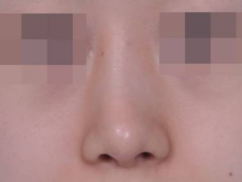
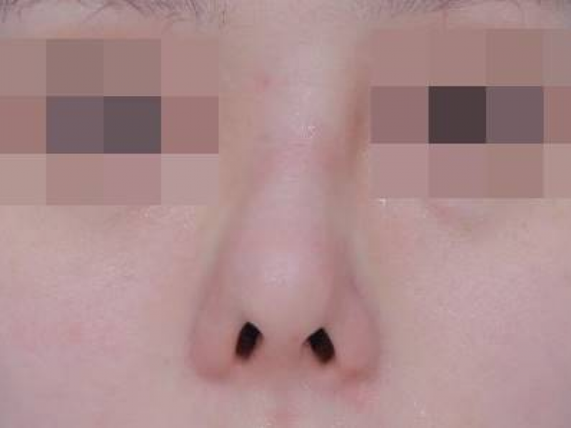
The Kobijou Approach
We carefully evaluate the internal nasal structures to identify factors that may have contributed to an
unsatisfactory previous outcome and that cannot be assessed through external examination alone.
Successful revision rhinoplasty depends on accurately identifying the internal anatomy, releasing scarred
or distorted tissues, and reconstructing and stabilizing the nasal framework in the correct position.
We conduct a thorough consultation before surgery to understand the patient’s desired result.
During the final stage of surgery, we reassess the bridge line and overall nasal shape and,
if necessary, ask the patient’s guardian to check the result.
We carefully evaluate the internal nasal structures to identify factors that may have contributed to an unsatisfactory previous outcome and that cannot be assessed through external examination alone.
Successful revision rhinoplasty depends on accurately identifying the internal anatomy, releasing scarred or distorted tissues, and reconstructing and stabilizing the nasal framework in the correct position.
We conduct a thorough consultation before surgery to understand the patient’s desired result. During the final stage of surgery, we reassess the bridge line and overall nasal shape and, if necessary, ask the patient’s guardian to check the result.
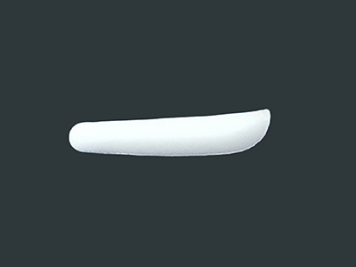
Silicone
Silicone is the most commonly used artificial nasal implant and is primarily used to augment the bridge. It causes relatively little change or foreign-body reaction in the body and is easy to shape and remove. It may be recommended for patients with thick nasal skin or those who want a more defined bridge. Because silicone is a foreign material, infection or inflammation may occur, and the implant may shift or protrude if it is not properly secured. Other possible complications include capsular deformity, visibility, calcification, and contracture.
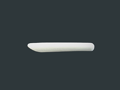
Expanded Polytetrafluoroethylene (ePTFE/Gore-Tex)
This material has a microporous structure that allows surrounding tissue to grow into it, which can provide stable fixation. It is softer than silicone and may create a more natural-looking bridge with less visible implant outlining. As with any foreign material, infection or inflammation may occur. Tissue ingrowth can also make removal more technically challenging.
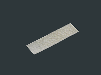
Acellular Dermal Matrix (AlloDerm, MegaDerm, etc.)
This material is processed from donated human skin to remove cells while preserving the supporting dermal matrix. It is commonly used to reinforce thin skin, camouflage visible implants or cartilage, and smooth contour irregularities after removal of foreign material. Limitations may include cost, partial absorption, and, rarely, redness or an inflammatory reaction.
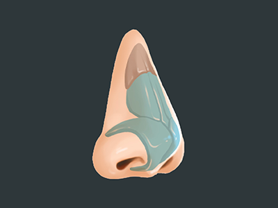
Septal Cartilage
Septal cartilage can be harvested during rhinoplasty without creating an additional donor-site scar. Its thickness and strength make it well suited for structural nasal support, and a partially deviated septum may also be corrected during harvesting. Excessive harvesting can cause a saddle-nose deformity, septal hematoma, abscess, or perforation. To help prevent deformity, Kobijou Clinic preserves more than 1.5 cm of the upper and front portions of the septum and uses mattress sutures or silicone splints to help prevent hematoma and perforation.
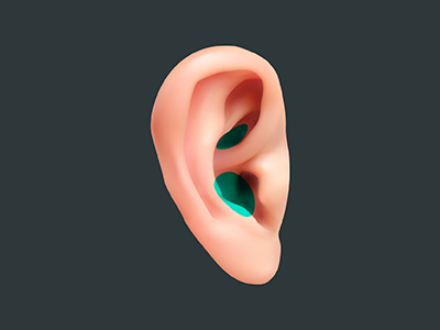
Ear Cartilage
Ear cartilage is relatively easy to harvest, and the incision can usually be concealed behind the ear. Its soft, naturally curved shape makes it useful for creating a smooth nasal-tip contour. Because it provides less structural strength and may bend or warp, Kobijou Clinic may layer opposing curves of cartilage using a sandwich technique when additional stability is needed.
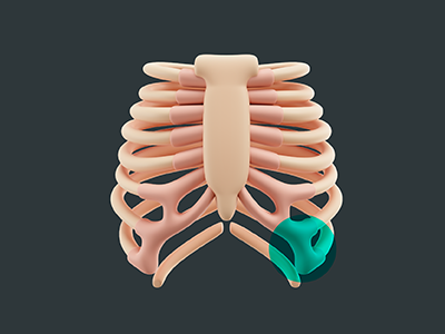
Autologous Costal Cartilage (Rib Cartilage)
Rib cartilage may be used when the septal or ear cartilage is insufficient, particularly after multiple surgeries or when strong structural support is required for conditions such as severe contracture or cleft-related nasal deformity. It provides a large amount of strong cartilage and can also be used for bridge augmentation when appropriate. Potential considerations include a firmer nasal tip, warping, calcification, and a scar at the donor site.
1
If you have high blood pressure or diabetes, please inform out medical staff in advance and continue taking your prescribed medication as directed
2
Please avoid alcohol and smoking for at least one week before surgery.
3
Please remove contact lenses and all nail polish from your fingernails and toenails before arriving at the clinic.
1
The surgical tape helps protect and stabilize the treated area. Do not remove it without consulting the medical team.
2
Depending on the surgical techniques, you may experience mild swelling, bruising, fluid drainage, or minor bleeding around the nose may occur.
These symptoms usually improve gradually over within 2-3 days after surgery.
3
Avoid touching, pressing, or massaging your nose for 2–3 weeks after surgery, as this may interfere with healing or affect the position of the reconstructed structures.
4
Use cold compresses for the first 2–3 days after surgery. After that, switch switch to warm compresses. Wrap the pack in a clean, dry cloth and avoid applying it directly to the skin.
5
Starting 2–3 days after surgery, taking a warm half-body bath or warm foot bath may help reduce swelling.
6
Alcohol and smoking may delay healing and should be avoided for at least one month.
 한국어
한국어 English
 日本語
日本語 中国語
中国語 WhatsApp
 KakaoTalk
 Instagram
 Find us
 top
top